Knee Muscle Anatomy Mri : Patellofemoral Problems | The Knee Doc / 4, infrapatellar fat pad of hoffa.
Knee Muscle Anatomy Mri : Patellofemoral Problems | The Knee Doc / 4, infrapatellar fat pad of hoffa.. General anatomy and musculoskeletal system. Involved early gray = muscle: Overuse injuries of the knee include tendonitis, bursitis, muscle strains, and iliotibial band syndrome. Want to learn more about it? This section of the website will explain.
Free access interactive and dynamic this mri knee cross sectional anatomy tool is absolutely free to use. Magnetic resonance imaging (mri scan): Click now to learn more about the bones, muscles, and soft tissues of these regions at leg and knee anatomy: Tips to keep joints healthy. Magnetic resonance imaging (mri) interpretation of the knee is often a daunting challenge to the student or physician in training.
Mri anatomy of knee Dr. Muhammad Bin Zulfiqar from image.slidesharecdn.com Injuries of the patellofemoral joint. Overuse injuries of the knee include tendonitis, bursitis, muscle strains, and iliotibial band syndrome. Want to learn more about it? Scroll through the structures to understand the anatomy. Magnetic resonance imaging (mri) is the test of choice to confirm the diagnosis of a torn meniscus. Support the body in an upright position without the need for muscles to work. The muscles that affect the knee's movement run along the thigh and calf. The muscles of the knee joint are incredibly important.
The knee is designed to fulfill a number of functions:
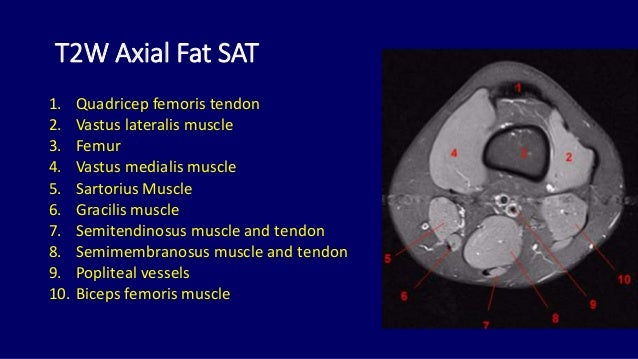
These muscles work in groups to flex, extend and stabilize the extending along the anterior surface of the thigh are the four muscles of the quadriceps femoris group (vastus lateralis, vastus medialis, vastus. Radiology imaging medical anatomy human anatomy and physiology anatomy study. Magnetic resonance imaging (mri) is the test of choice to confirm the diagnosis of a torn meniscus. Support the body in an upright position without the need for muscles to work. Knee anatomy francesc malagelada jordi vega pau golanó the knee is the largest joint in the human body and one of the most complex from a functional point of view. This mri knee cross sectional anatomy tool is absolutely free to use. Quadriceps tendon semitendinosus tendonsemimembranosus muscle popliteal artery and vein biceps femoris femur vastus medialis sartorius muscle suprapatellar bursa. Click now to learn more about the bones, muscles, and soft tissues of these regions at leg and knee anatomy: These are essential structures to evaluate in routine assessment of the knee on mri. Injuries of the patellofemoral joint. It is a noninvasive test that can visualize the inner structures of the knee, including the cartilage and ligaments, the surface of the bones, and the muscles and tendons that surround the knee joint. The knee is designed to fulfill a number of functions: Magnetic resonance imaging (mri scan):
Rubin da, kettering jm, towers jd, britton ca: It is also one of the most often injured joints because of its anatomic characteristics, the interrelation of its structural components. This mri knee cross sectional anatomy tool is absolutely free to use. Master leg and knee anatomy using our topic page. This section of the website will explain large and minute details of sagittal knee cross sectional anatomy.
Mri anatomy of knee Dr. Muhammad Bin Zulfiqar from image.slidesharecdn.com Scroll through the structures to understand the anatomy. General anatomy and musculoskeletal system. An understanding of normal anatomy and biomechanics of the knee extensor mechanism is necessary to comprehend the imaging of extensor mechanism injuries. Want to learn more about it? This section of the website will explain. Mr imaging of knees having isolated and combined ligament injuries. Radiology imaging medical anatomy human anatomy and physiology anatomy study. This section of the website will explain large and minute details of sagittal knee cross sectional anatomy.
Use the checklist to quiz yourself.
General anatomy and musculoskeletal system. Click on the links to show each structure. Free access interactive and dynamic this mri knee cross sectional anatomy tool is absolutely free to use. Tips to keep joints healthy. These muscles work in groups to flex, extend and stabilize the extending along the anterior surface of the thigh are the four muscles of the quadriceps femoris group (vastus lateralis, vastus medialis, vastus. Use the checklist to quiz yourself. This section of the website will explain. An understanding of normal anatomy and biomechanics of the knee extensor mechanism is necessary to comprehend the imaging of extensor mechanism injuries. Rubin da, kettering jm, towers jd, britton ca: Mri for evaluating knee pain in older patients: Magnetic resonance imaging (mri) interpretation of the knee is often a daunting challenge to the student or physician in training. This mri knee cross sectional anatomy tool is absolutely free to use. They are attached to the femur (thighbone), tibia (shinbone), and fibula (calf bone) by fibrous tissues called ligaments.
This section of the website will explain. Magnetic resonance imaging (mri) is the test of choice to confirm the diagnosis of a torn meniscus. Want to learn more about it? General anatomy and musculoskeletal system. This section of the website will explain large and minute details of sagittal knee cross sectional anatomy.
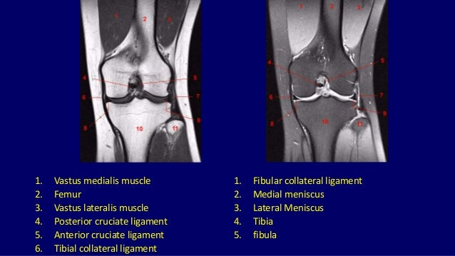
Knee MRI Scan, Sagittal from ar.utmb.edu Click now to learn more about the bones, muscles, and soft tissues of these regions at leg and knee anatomy: This webpage presents the anatomical structures found on knee mri. Each anatomical structure was labeled interactively. Mri for evaluating knee pain in older patients: The muscles of the knee include the quadriceps, hamstrings, and the muscles of the calf. Tendons attach the muscles to each other. Mri anatomy and positioning series module 2: The journal of musculoskeletal medicine.
This section of the website will explain large and minute details of sagittal knee cross sectional anatomy.
This section of the website will explain. Mri anatomy and positioning series module 2: The knee is designed to fulfill a number of functions: This section of the website will explain large and minute details of sagittal knee cross sectional anatomy. This webpage presents the anatomical structures found on knee mri. Learn about mri anatomy with free interactive flashcards. Each anatomical structure was labeled interactively. Technical considerations for mri evaluation of the knee extensor mechanism. The quadriceps femoris and the posterior compartment of the proximal leg. Injuries of the patellofemoral joint. 4, infrapatellar fat pad of hoffa. Muscles in the posterior compartment of the thigh. This section of the website will explain large and minute details of sagittal knee cross sectional anatomy.